
I posted this lesion earlier, this is a look from a different angle to make a point about a couple of diagnostic algorithms.
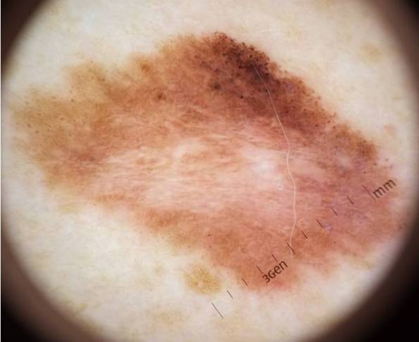
If we apply the 2 step algorithm, we find
- it is melanocytic, because we see reticular network, also brown globules, either of which are characteristic of melanocytic naevi. Also, it is melanocytic by default since there are no clues allowing us to positively identify another type of skin lesion, e.g. seborrhoiec keratosis, haemangioma, BCC etc.
- if melanocytic (which it is-see above) could it be a melanoma? Or to ask the same question in a different way, can we say with confidence that it is benign? The answer is, no we can’t prove its benign because it is very irregular-both pattern and colour evidence significant lack of symmetry.
We must therefore excise as a suspected melanoma.
If we apply the Harald Kittler ‘chaos and clues’ algorithm we get the same answer by a different route.
- Is the lesion chaotic? (Kittler defines chaotic as more than one pattern and/or more than one colour. However, there are degrees of chaos between zero and complete and utter chaos, we get a feel for evaluating degrees of chaos by studying hundreds of lesions). Anyway, we see structureless areas plus lines reticular and clods, also at least 3 colours, so yes it’s chaotic.
- Since chaotic, look for clues. I have already mentioned clues to melanocytic, and no clues to anything else, so by the Kittler method we now have a chaotic melanocytic lesion, which by definition must be excised.
Histology confirmed a thin (therefore eminently curable) melanoma.

The following text has been copied from an item in the Worksop Guardian (link below) which came up on my Google alert on melanoma.
>>>>>>>>>John Mann MP has launched a campaign against skin cancer in Bassetlaw after it emerged that the area has the highest number of people suffering from melanoma in the country.
Melanoma is the UK’s fifth most common cancer (*) with rates increasing more rapidly than any of the current ten most common cancers for both men and women.
Mr Mann said: “It is of huge concern that so many people in Bassetlaw suffer from this type of skin cancer and it’s important that patients are aware of the dangers of melanoma and for patients who receive an advanced diagnosis to be able to access a number of treatments available.
“I will be working with the charity Melanoma UK to raise awareness of this cancer in Bassetlaw and supporting their work to ensure appropriate treatments are available on the NHS.
“Anyone who sees any sign of skin changes such as bleeding moles should play it safe and immediately make an appointment to see their GP. The NHS website has further advice on symptoms and treatment.” (my emphasis-SH)
Gillian Nuttall, founder of Melanoma UK, said: “I am delighted that John Mann MP has launched a campaign to raise more awareness of melanoma in Bassetlaw. Treated early, melanoma is one of the most curable cancers; left late, it is one of the most deadly.” <<<<<<<<<<<<<
Now this is all very well, but it mainly seems to me to be about lobbying for the very expensive new palliative drugs for stage 4 melanoma (I won’t name them but they can be easily Googled). These drugs are horrifically expensive (theoretically up to half a million pounds per patient for combined and repeated courses) have severe (sometimes fatal) side effects and so far only a few patients have survived more than 2 years. The benefits are being played up and the down side played down. I’m not saying these drugs shouldn’t be used, just that we must have a calm debate about the costs, risks and (limited) benefits and not simply say ‘The NHS must fund them, fullstop!’
All this is debatable and others’ views will not be the same as mine. But my main problem with the statement above is that it gives worse than useless advice about early diagnosis, which we know saves lives.
The statement that people with ‘bleeding moles’ should see their GP to ‘play it safe’ reveals a PROFOUND AND DANGEROUS IGNORANCE about the early diagnosis of melanoma. Friends, if you are unlucky enough to have a melanoma, which 1 in 50 of us over a lifetime will be, then by the time it is BLEEDING your chances of survival are much reduced. Most skin lesions that bleed will be benign (e.g. traumatised wart) or low grade cancer (e.g. basal cell cancer which is 99% curable). most melanomas will look abnormal for months to a year or more before they start bleeding. Bleeding is a LATE sign of ADVANCED melanoma.
The whole point of dermoscopy, and of this blog and my course, is to pick up melanomas at a thin and CURABLE stage well before they have grown big and thick enough to start bleeding.
Gillian Nuttall’s comment is more sensible but does not go far enough (fair enough she may have said more that wasn’t reported). We need a trained dermoscopist in every health centre to competently check every mole that is changing colour, shape or size or which just looks wrong.
Since melanoma is now killing more than twice as many people as cancer of the cervix, for which we have universal screening and now a vaccine, why on earth not? The ‘new treatments’ that The Honourable Mr Mann MP is advocating for will, as I mentioned in an earlier post, cost the NHS £billions, and they are for the most part only palliative-giving an extra 6 or 12 months of survival. We should be putting a much bigger effort into earlier diagnosis and dermoscopy training for GPs and practice nurses is key to this. (**)
The drug companies and advanced melanoma sufferers are vocally advocating for the NHS to find much new money to fund these VERY costly drugs, but who is advocating for earlier diagnosis, which we know saves more lives and saves them much cheaper?
(*) only if you don’t count non melanoma skin cancer, which is far and away our most common cancer. Usually omitted from statistics as it is rarely fatal.
(**) declaration of interest, SH earns reward for providing this kind of education and training.
I receive a regular Google Alert on melanoma 3 or 4 times a week. Much of the information concerns new medicines for metastatic melanoma and the biotech and pharma companies that make and sell them. Other alerts concern the stories of individual melanoma patients, fundraising etc. One story that comes up a lot is smartphone apps that can apparently help with earlier diagnosis. There are now dozens of these apps.
The latest app to come to my attention in Skinvision. See here, there is a link to a video.
I have not done an appraisal of these apps, and I am neither for nor against them. Some work on mole recognition software, others send images to experts who give an opinion, some link to educational images on line, others enable people to photograph, store images of and follow their own moles digitally (the Skinvision does several of these things according to the video).
None of this is wrong in principle, but as ever its the detail and the actual performance in real life that counts-we ought to be skeptical and watch out for unintended effects and pitfalls of new medical applications-good intentions are not enough, the thing has to WORK, and also be safe. One thing which worries me is the possibility that some of these folks behind these devices might be selling their services as mole removal surgeons and therefore IN THEORY might have a financial interest in diagnosing lots of moles as potentially worrying and needing surgical removal (ker-ching!) {*}
The efficacy and safety of Skinvision and other similar apps would have to be determined by an appropriate professional body or investigator. In principle, they might be a very good thing, but there are drawbacks with any diagnostic system, whether through over diagnosis (false positives) or under diagnosis (false negatives). Anyone involved in cancer diagnosis is terrified of a false negative (people can die from missed melanomas and even if there was no higher motive, doctors are afraid of getting sued in this event). There is therefore a tendency of diagnostic systems to over diagnose, and this may lead to anxiety and avoidable excisions. Just saying.
But we do know there is are potential benefits of skin monitoring. At the 4th World Dermoscopy and Skin Imaging Congress in Vienna last April, I heard 2 presentations from top world experts about the benefits of digitally monitoring the skin for suspicious moles. Professor Scott Menzies of Sydney, Australia presented about short and long term digital monitoring, where high risk patients had quality digital images of their whole skin and/or suspicious individual moles made and were followed up. Basically, if a mole did not change over time, it was OK. If it did change (size, shape, colour, new appearance) it was more carefully evaluated and if in doubt removed. This proved to be cost effective (avoided unnecessary excisions of marginally worrying moles) and safe (the melanomas detected and removed by this system were thin and easily curable).
So, we have good evidence that professional digital monitoring of high risk patients and worrying flat (never thick) lesions is safe, effective and saves people having operations and scars they don’t need. Sounds like a win win. But setting this up and running it costs money and is only cost effective for high risk patients (e.g. people with 100 or more moles and a history of melanoma). But what if we could use an inexpensive app and a camera or tablet/smartphone we already have to do it ourselves? Not as good as an expert, perhaps, but the principle is the same and, as Bluto famously said in ‘Animal House’, ‘Don’t cost nothing!’
Another talk was by Professor Peter H Soyer who presented some research about people using a mini dermoscope that attached to their mobile phone to photograph any mole they were worried about and email the image to the dermatologist. The evaluation of the emailed mole image compared with face to face consultation very favourably. This was not a proper trial but a test of concept-certainly it seems to offer potential. This paper was published in April 2015’s British Journal of Dermatology.
So, where does that leave us for now? The gold standard of digital monitoring with a dermatologist and state of the art equipment is not achievable for all, it remains the preserve of the wealthy worried well or high risk patients in enlightened and adequately funded health care systems (so not the British NHS then). But most of us in wealthy countries have access to a smartphone or tablet.
In a future post I will consider how individuals who are worried but don’t have easy access to a dermatologist can do their own mole mapping with an Ipad or similar. Basically, photograph the whole of your skin, or as much as modesty allows (wear a bikini) and check your skin against it every 3 months or so. Be reassured if nothing changes. If of course you do get something new or changing then you have evidence you can take to your GP for onward referral to a specialist.
I must stress that self photography of the skin and home monitoring is not as yet evidence proven by proper trials, but it seems harmless and makes sense. Think of it as ‘poor man’s digital monitoring’ and certainly top world melanoma expert Scott Menzies has shown that monitoring can relieve anxiety and save lives.
{*} for younger non British readers, the word ‘ker-ching!’ is meant to sound like an old fashioned cash register ringing up a sale.
I edit dermoscopic images, using Microsoft photo editor. Others do as well. this isn’t cheating, after all the image we take with a digital camera in only a representation of the natural features of the lesion, and may not be a true representation.
Obviously photoshopping in features which aren’t there would not be legitimate, but I often edit images for brightness and contrast which makes them easier to see key features like network and vessels.

Here is a seborrhoiec wart, poor image quality. Now here it is again, edited.

the brown clods and white clods are easier to see.
Here is another from the same patient.


No cheating, just clarification. One of the presenters at the Vienna world congress on dermoscopy and skin imaging made a similar point. It is worth trying. also it’s often a good idea to crop images.
Any clinician who does dermoscopy should consider image capture and storage (remember patient confidentiality especially if recognisable features are included). benefits include personal audit and the ability to teach others, we need more experienced dermoscopy teachers.
This is a common presentation, someone notices a funny looking mole on their own or someone else’s back and suggests they get it checked. Seems reasonable, after all melanoma skin cancer is on the increase and it can kill you.

This person has a dozen or so small harmless looking ‘moles’ on their upper back, and one that sticks out like a sore thumb, or ugly duckling. Closer evaluation is required.

Mostly quite featureless. certainly no features such as reticular network or brown globules to make you think of a melanocytic lesion. There are several brownish clods of variable sizes-these are the comedo like openings of seborrhoiec keeratosis. How do we tell these from globules? Study hundreds of images-they are different.
Closer examination of the almost (but not quite ) featureless background reveals many small homogenous blood vessels. The pattern is small loops, typical of seborrhoeic keratosis. Which is the diagnosis. Other supporting features of seb k are the very well defined edge and fissures. an autocorrected image shows these fissures (circled) more readily. Click on the image to enlarge.

There is a blood vessel structure in the upper right quarter near the centre, the circle goes through it. This is just random. How can I say that with confidence? By studying many thousands of lesions. There are plenty of features here to support seb k, no features of skin cancer, and we simply do see varied random features in all kinds of skin lesions.
Many ugly ducklings are harmless, and can be proved so with the dermoscope, saving time, money and worry.
